- Introduction
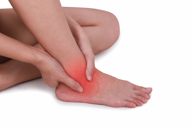
The ankle joint is a complex, weight-bearing structure prone to injury and degeneration due to its constant use in daily activities and sports. When conservative treatments fail to relieve chronic pain, stiffness, or instability, ankle arthroscopy may be recommended.
Ankle arthroscopy is a minimally invasive surgical procedure that allows orthopedic surgeons to examine and treat various ankle conditions using a small camera and instruments inserted through tiny incisions. It can be used for diagnosis, debridement, ligament repair, removal of loose bodies, cartilage restoration, and synovectomy, among others.
This comprehensive guide will help you understand the anatomy of the ankle, indications for arthroscopy, the procedure itself, recovery, risks, and advancements in technology and technique.
- Ankle Joint Anatomy
The ankle is a hinge joint, formally known as the tibiotalar joint, composed of three bones:
- Tibia (shin bone)
- Fibula (outer lower leg bone)
- Talus (uppermost foot bone)
These bones are stabilized by ligaments, tendons, and cartilage, all of which may be addressed during arthroscopy.
Key Supporting Structures:
- Medial and lateral collateral ligaments
- Deltoid ligament complex
- Syndesmosis: holds the tibia and fibula together
- Articular cartilage: lines joint surfaces for smooth movement
- Subtalar joint: below the ankle, allows inversion and eversion
- Common Indications for Ankle Arthroscopy
Ankle arthroscopy is used to diagnose and treat a wide variety of intra-articular conditions. Some of the most common include:
- Osteochondral Lesions of the Talus (OLT)
- Cartilage and bone injury on the top of the talus bone
- Often due to ankle sprains or trauma
- Causes chronic pain, swelling, and catching
- Synovitis
- Inflammation of the joint lining
- May be due to injury, overuse, or autoimmune diseases
- Anterior Ankle Impingement ("Footballer’s Ankle")
- Bony spurs or soft tissue that cause pain during dorsiflexion
- Loose Bodies
- Fragments of bone or cartilage that float within the joint
- Lead to locking, catching, or pain
- Chronic Ankle Instability
- Caused by recurrent sprains and ligament damage
- Arthroscopy may be used to evaluate and assist in ligament reconstruction
- Post-Traumatic Arthritis
- Inflammation and degeneration of cartilage after injury
- May require debridement or microfracture
- Infection / Septic Arthritis
- Though rare, arthroscopy is sometimes needed to clean infected joints
✅ Note: Ankle arthroscopy can also assist in fracture management, tendon evaluation, and even subtalar pathology.
- Diagnostic Process: When is Arthroscopy Recommended?
Before arthroscopy is recommended, a complete evaluation is performed, including:
- History and Physical Exam
- Swelling, pain location, instability, ROM limitations
- X-rays
- To evaluate bony anatomy, alignment, and arthritis
- MRI
- Best for viewing cartilage lesions, ligaments, and soft tissue
- CT scan
- Helpful for evaluating bone detail in osteochondral lesions
- Diagnostic injection
- Anesthetic is injected to determine if pain comes from the joint
Surgery is typically considered when non-surgical options (e.g., rest, NSAIDs, bracing, physical therapy, injections) have failed for 3–6 months or more.
- Ankle Arthroscopy Procedure: Step-by-Step
Ankle arthroscopy is generally performed under general, spinal, or regional anesthesia and takes about 30 to 90 minutes, depending on the complexity of the case.
🛏️ Step 1: Patient Positioning
- Patient is placed supine (on their back) or prone (on their stomach) depending on the target location inside the ankle.
- A tourniquet is applied to minimize bleeding for better visibility.
🔧 Step 2: Portal Placement
- Two to three small incisions (portals) are made—typically at the front (anterior) or back (posterior) of the ankle.
- A cannula and arthroscope (tiny camera) are inserted to visualize the joint.
🧼 Step 3: Diagnostic Arthroscopy
- The surgeon inspects:
- Articular cartilage
- Synovium
- Ligaments
- Bone surfaces
- Presence of loose bodies or scar tissue
🛠️ Step 4: Therapeutic Procedures (as needed)
- Debridement: Trimming of damaged cartilage or scar tissue
- Synovectomy: Removal of inflamed synovial tissue
- Microfracture: For cartilage lesions—drilling small holes into bone to stimulate healing
- Removal of loose bodies or bone spurs
- Assistance with ligament repair in cases of chronic instability
🔚 Step 5: Closure and Post-op Dressing
- Portals are closed with sutures or steri-strips.
- A sterile dressing and compression wrap is applied.
- Some patients may be placed in a splint or boot.
🕒 Duration: 30–90 minutes depending on pathology
🏥 Outpatient Procedure: Most patients go home the same day
- Postoperative Recovery and Rehabilitation
Recovery from ankle arthroscopy varies depending on what was performed during surgery (simple debridement vs. microfracture vs. ligament repair).
📆 General Recovery Timeline:
|
Phase |
Timeframe |
Focus |
|
Phase 1 |
0–2 weeks |
Pain/swelling control, limited weight-bearing |
|
Phase 2 |
2–6 weeks |
Gradual weight-bearing, early ROM exercises |
|
Phase 3 |
6–12 weeks |
Strengthening, proprioception, balance training |
|
Phase 4 |
3–6 months |
Return to sport, advanced conditioning |
🦶 Weight-Bearing Guidelines:
- Simple debridement/synovectomy: Weight-bearing as tolerated in 1–3 days
- Microfracture/cartilage repair: Non-weight-bearing for 4–6 weeks
- Ligament repair: May require immobilization and delayed weight-bearing for 4–6 weeks
💪 Physical Therapy Components:
- Ankle pumps, isometric exercises, towel stretches
- Resistance bands for dorsiflexion/plantarflexion
- Balance board drills, proprioceptive training
- Sport-specific reconditioning
💡 Key to Success: Adherence to rehab protocol is essential to regain strength, prevent stiffness, and reduce the risk of complications.
- Risks and Complications
Although ankle arthroscopy is considered a safe and minimally invasive procedure, it still carries some risks — especially when performed for complex pathology or in anatomically tight joints like the ankle.
⚠️ Common Risks:
- Swelling and bruising (temporary)
- Joint stiffness: May occur if rehabilitation is delayed or too aggressive
- Infection: Rare, but can occur at portal sites or inside the joint
- Blood clots (DVT): Especially in patients with risk factors (e.g., smoking, immobility)
- Painful scar tissue or adhesions
⚙️ Procedure-Specific Complications:
- Nerve injury: Due to proximity of nerves (superficial peroneal, sural, or saphenous) to the portal sites; can lead to numbness, tingling, or nerve pain
- Bleeding or hemarthrosis: Blood accumulation in the joint
- Instrument breakage: Extremely rare with modern technology
- Delayed healing: Especially after microfracture or cartilage work
🔍 Complication rates are low (~5–8%) when performed by experienced orthopedic surgeons. Most issues are mild and resolve with proper care.
- Success Rates and Long-Term Outcomes
Ankle arthroscopy has shown excellent outcomes for a variety of pathologies when paired with appropriate surgical indications and a structured rehabilitation program.
📈 Success Rates by Condition:
|
Condition |
Success Rate |
|
Synovitis / Impingement |
85–95% |
|
Loose body removal |
90–95% |
|
Osteochondral lesion (OLT) |
70–85% (microfracture) |
|
Ligament repair + arthroscopy |
80–90% |
|
Early arthritis debridement |
60–75% (temporary relief) |
⏳ Long-Term Outcomes:
- Most patients report sustained pain relief and improved mobility for 5–10 years, depending on activity level and underlying joint condition.
- Athletes can return to their sport in 3–6 months, depending on the procedure.
- Reoperation rates are low but may increase if arthritis progresses or if instability isn’t addressed.
🧠 Patient satisfaction is generally high, especially in cases where other conservative treatments have failed.
- Innovations in Ankle Arthroscopy
Ankle arthroscopy continues to evolve with new tools, techniques, and biological enhancements aimed at improving outcomes and reducing recovery times.
🧬 Biologic Therapies
- Platelet-Rich Plasma (PRP): Used intraoperatively or post-op to enhance healing in cartilage or ligament procedures.
- Stem Cell Injections: Emerging role in regenerating cartilage and modulating inflammation, especially for osteochondral lesions.
🦴 Advanced Cartilage Repair Techniques
- Microfracture Plus (augmented microfracture): Combines microfracture with collagen scaffolds to support better cartilage regeneration.
- Autologous chondrocyte implantation (ACI): Harvesting and culturing the patient’s own cartilage cells, though still rare in ankle joints.
🤖 3D Imaging and Navigation
- Use of preoperative CT or MRI mapping to precisely target lesions
- Helps in tailoring portal placement and planning complex reconstructions
🪡 Minimally Invasive Ligament Repair Systems
- Devices like InternalBrace™ and Broström-Gould modifications enable reinforced repairs with smaller incisions and earlier mobilization
🧠 Artificial Intelligence (AI) in Preoperative Assessment
- AI tools are being developed to assist in predicting surgical outcomes and tracking joint degeneration using MRI data.
🚀 These innovations aim to reduce trauma, enhance healing, and offer more personalized care based on joint pathology and patient lifestyle.
- Patient FAQs
❓ Is ankle arthroscopy painful?
- Some discomfort is expected, especially in the first week. However, pain is usually manageable with medications, ice, and rest.
❓ How long will I be on crutches?
- It depends on the procedure. For simple debridement, you may walk within days. For microfracture or ligament repair, crutches are required for 4–6 weeks.
❓ When can I return to sports?
- Typically 3–6 months, depending on procedure complexity and rehab progress.
❓ Will I need physical therapy?
- Yes. A structured rehab program is essential to restore strength, range of motion, and proprioception.
❓ Are the incisions visible?
- Portals are small (typically 5–10 mm) and heal with minimal scarring. They’re usually placed in low-visibility areas.
❓ Is arthroscopy better than open surgery?
- For many conditions, yes—it offers faster recovery, less pain, and better visualization with less soft tissue damage. However, not all problems can be solved arthroscopically.
❓ Will the problem come back?
- If the root cause (e.g., instability, arthritis, poor mechanics) isn’t addressed, symptoms may recur. Following rehab protocols and modifying risk factors is key.


Dodajte Komentar